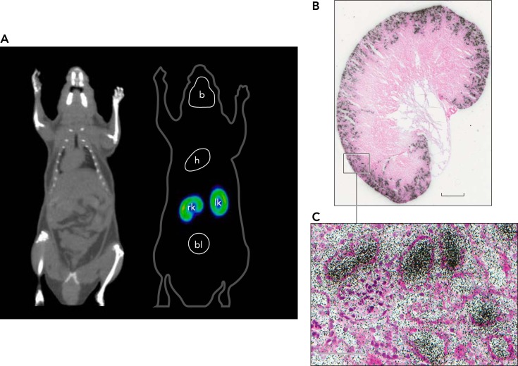
FIGURE 3.
Location of SGLT2 in a mouse using [18F]-dapagliflozin micro-PET and ex vivo autoradiography
A: dapagliflozin, a selective high-affinity SGLT2 inhibitor (Kd 2 nM) labeled with [18F], was injected intravenously into a mouse, and the distribution was recorded using micro-PET (16). Binding was only observed in the outer cortex of the kidney (bright yellow band), and this was rapidly displaced with intravenous injection of dapagliflozin or phlorizin (not shown). No binding was observed in Sglt2-null mice. B: after [18F]-dapagliflozin binding to the kidney reached a steady state (15 min), a kidney was rapidly removed and prepared for ex vivo autoradiography. A slice of the whole kidney shows [18F]-dapagliflozin binding to the outer cortex, and a higher magnification view shows the inhibitor within some tubules adjacent to the glomerulus. Redrawn from Ref. 18.

