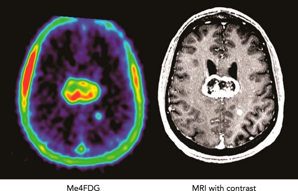
FIGURE 4.
SGLT activity in brain cancer patients using Me-4FDG PET
Me-4FDG PET scans and a contrast MRI were conducted on a patient with a grade IV astrocytoma in the corpus callosum. The co-registered images of the brain show uptake of Me-4FDG and the contrast agent into a large 46-mm tumor with a non-enhancing necrotic core in the corpus callosum and a small 6-mm tumor in the left parietal gray matter. The PET signal was approximately twice as high as that in the blood in the sagittal sinus, and the signal-to-noise ratio for the tumor relative to the rest of the brain was 12. See Ref. 24.

