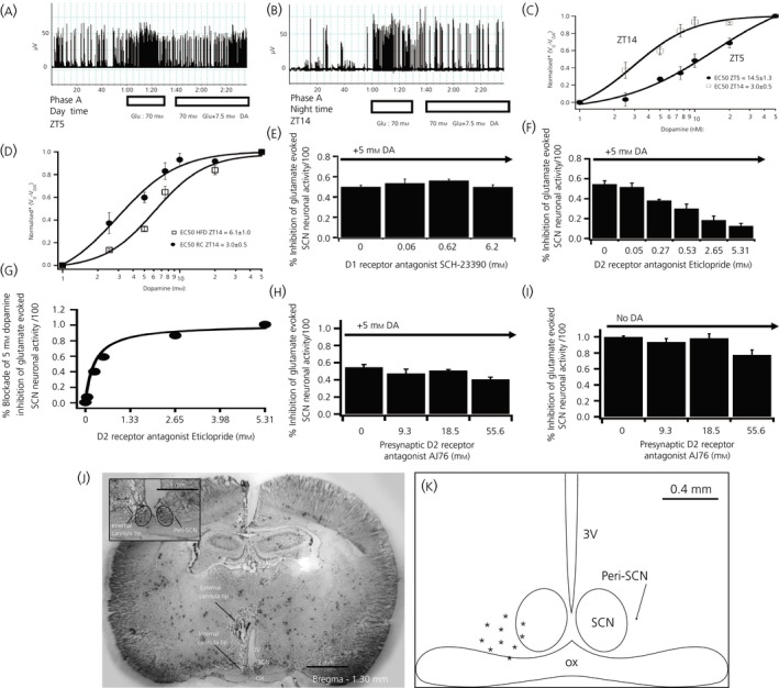
Figure 4.

Daily peak of suprachiasmatic nuclei (SCN) neuronal electrophysiological response to dopamine coincides with the circadian peak of dopamine release at SCN in healthy insulin‐sensitive animals and is reduced in high‐fat diet (HFD) fed rats. (A, B) Electrophysiological recording example of dopamine (DA) (7.5 mM) inhibition of glutamate (70 mM) evoked SCN neuronal activity at Zeitgeber time (ZT)5 (day time) and at ZT14 (just at darkness onset and the onset of locomotor activity in these nocturnal rodents). (C) Daily variation of peri‐SCN/SCN electrophysiological sensitivity to peri‐SCN dopamine inhibition (1, 2.5, 5, 10, 25 and 50 mm) of glutamate stimulation of SCN neurones at ZT5 and ZT14. A two‐way anova with repeated measures indicates a significant difference between the SCN neuronal response to dopamine inhibition at ZT14 and at ZT5 (F 1,35 = 25.597, P < .001). The half maximal effective concentration of dopamine inhibition to the SCN neuronal response at ZT5 (EC 50 = 14.5 ± 1.3 mm) was 4.8 times greater than at ZT14 (EC 50 = 3.0 ± 0.5 mm, P < .05). (D) SCN neuronal sensitivity response to peri‐SCN dopamine inhibition of peri‐SCN glutamate stimulation of SCN neurones at ZT14 in HFD and rodent chow (RC) fed rats. A two‐way anova with repeated measures indicates a much reduced SCN dopamine responsiveness at ZT14 in rats made obese by HFD feeding (F 6,48 = 54.3, P < .0001). (E) SCN neuronal responsiveness to dopamine inhibition in the presence of D1 specific receptor antagonist (SCH‐23390) at ZT14. SCH‐23390 did not alter peri‐SCN glutamate evoked SCN neuronal activity responsiveness to peri‐SCN dopamine inhibition at the tested dosages. (F,G) Glutamate‐evoked SCN neuronal activity responsiveness to peri‐SCN dopamine inhibition in the presence of D2 specific receptor antagonist (Eticlopride) at ZT14. D2 antagonist dose‐dependently blocked glutamate‐evoked SCN response to peri‐SCN dopamine (P < .05, One way anova). D2 antagonist eticlopride EC 50 = 265 μm. (H,I) Glutamate‐evoked SCN neuronal responsiveness in the presence of presynaptic antagonist (AJ76) at ZT14 with (H) or without (I) peri‐SCN dopamine applied. All values are the mean ± SEM (n = 5 per group). Higher dose AJ76 resulted in the slight inhibition of glutamate stimulation as may be expected from its potential consequent increase of endogenous synaptic dopamine level (subsequent to presynaptic dopamine D2 receptor blockade). (J) Histology of peri‐SCN injection cannula placement. The external cannula tip location was at the stereotaxic coordinates of anteroposterior −1.3, mediolateral 0.4 mm and dorsoventral −7.3 mm from dura. The injection cannula protruded 2 mm from the guide external cannula. The internal cannula tip shows the injection site. OX, optic chiasm; 3V, third ventricle. Enlarged peri‐SCN injection site is shown in the insert. The cannula placement was the same as the microdialysis probe location. (K) Diagrammatic representation of the peri‐SCN injection target site locations for animal brains confirmed histologically. The injection sites are between −0.1 mm and 0.4 mm lateral to the SCN. Only data from properly targeted cannula and probes were used for analyses. *Normalised V 0/VDA represents the fractional inhibition of glutamate evoked SCN neuronal activity relative to pre‐glutamate administration baseline activity level
