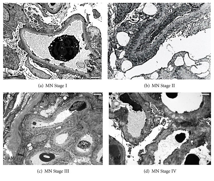
Figure 1.
Electron microscopy representative image of glomerular membranous nephropathy in adults. (a) Stage I: electron dense deposits, irregularly distributed, at the outside of the glomerular basement membrane (GBM), without inflammatory reaction around the deposits. There is a variable degree of foot process effacement. (b) Stage II: spikes are irregular projections of the GBM among the subepithelial deposits. (c) Stage III: with progression of the disease, spikes become longer and incorporate the deposits in a thickened GBM. (d) Stage IV: the deposits lose their electron density until disappearance in the advanced stages of the process. Magnification ×3000. Kindly provided by Jean Michel Goujon, M.D., Ph.D. (Centre National de Référence Maladies Rares: Amylose AL et Autres Maladies à Dépôts d'Immunoglobulines Monoclonales, Université de Poitiers; Pathology Department, Centre Hospitalier Universitaire de Poitiers, Poitiers, France).

