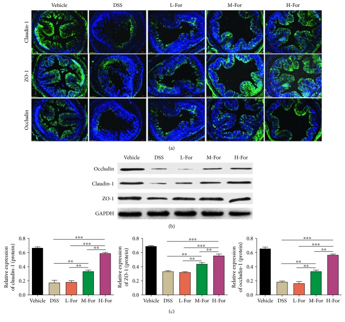
Figure 3.
For relieved DSS-induced colonic epithelial tight junction destruction in mice. (a) Representative immunofluorescence images for claudin-1 (Green), occludin (Green), and ZO-1 (Green) in the colonic tissue. (b, c) Protein levels of claudin-1, occludin, and ZO-1 in the colon tissues were analyzed by western blotting. ∗∗ p < 0.01, and ∗∗∗ p < 0.001.

