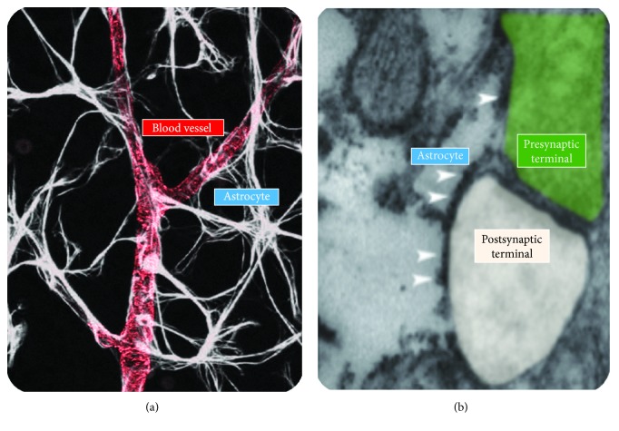
Figure 1.
Astrocytic interactions with blood vessels and synapses. (a) GFAP labeling of astrocytes (white) making contact with blood capillaries, visualized using antibodies directed against the smooth muscle-specific α-actin (ASMA) (red). Modified with permissions from [126]. (b) Transmitted electron micrograph of an excitatory synapse in the hippocampal area CA3, contacted by an astrocytic process. Arrowheads show PAR1 localization along the astrocytic membrane. Modified with permissions from [80].

