Figure 3.
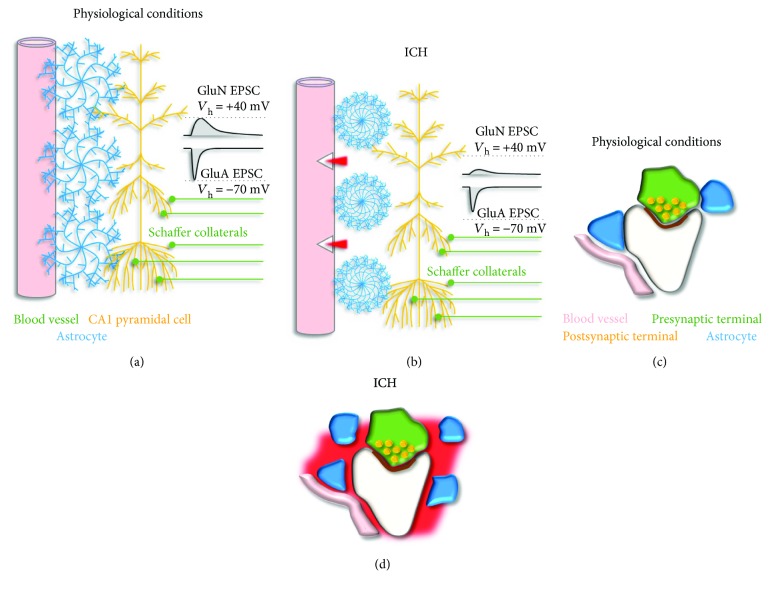
The coupling of astrocytes with blood vessels and synapses changes during ICH. (a) Schematic representation of the organization of blood vessels (pink), astrocytes (blue), and pyramidal cells in the hippocampal area CA1 (yellow) in physiological conditions. Astrocytes closely interact with both blood vessels and neurons. CA1 pyramidal cells receive excitatory afferents from area CA3 via the Schaffer collateral pathway (green). GluA and GluN receptors are expressed postsynaptically at Schaffer collateral synapses. Current responses mediated by GluA and GluN receptors can be isolated using pharmacological approaches and whole-cell patch-clamp recordings in voltage-clamp mode holding pyramidal cells at −70 mV or +40 mV, respectively (black). (b) Schematic representation of the structural reorganization of astrocytes in response to ICH and PAR1 activation. Astrocytic processes shrink, proliferate, and migrate further away from excitatory synapses. These changes cause faster glutamate clearance and weaken GluA- and GluN-mediated synaptic transmission [87]. (c) Schematic representation of the synaptic and perisynaptic environment in physiological conditions. (d) Schematic representation of the synaptic and perisynaptic environment in ICH. The remodeling of the neuropil induced by ICH is as described in (b).

