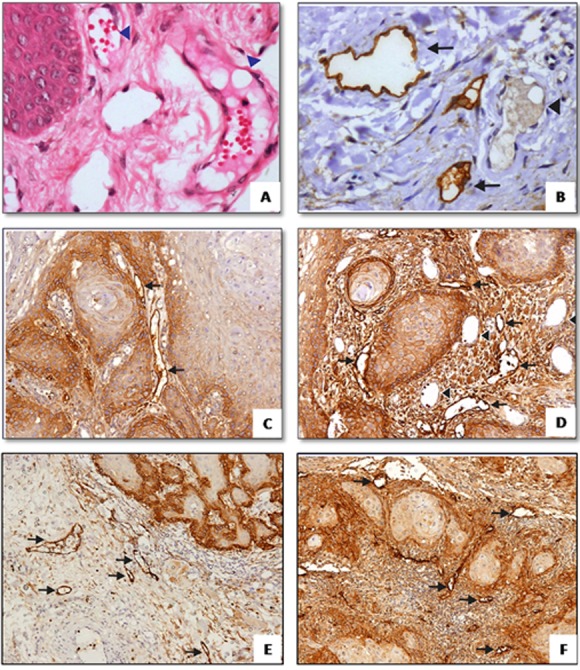
Figure1.

a: H&E staining of normal mucosa showing blood vessels (arrowhead) (400X). b: D2-40 immunostaining of normal mucosa, lymphatics are positively stained (arrow) and distinguished from blood vessels which are negatively stained (400X). D2-40 immunostaining of intratumoral lymphatics in superficial c and d: and deep, e and f: region (100X)
