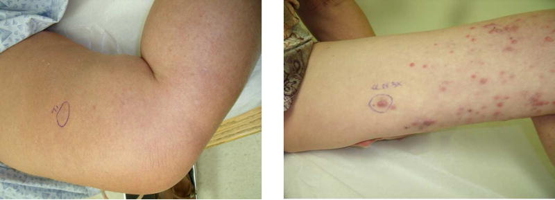
Figure 1.
A local recurrence (left). This single lesion is mapped using methods described plus vital blue dye as resection of the lesion is planned with SLNB. (Right) A patient with unresectable in-transit disease. The most proximal lesion (circled) is mapped using described methods and no blue dye is used.

