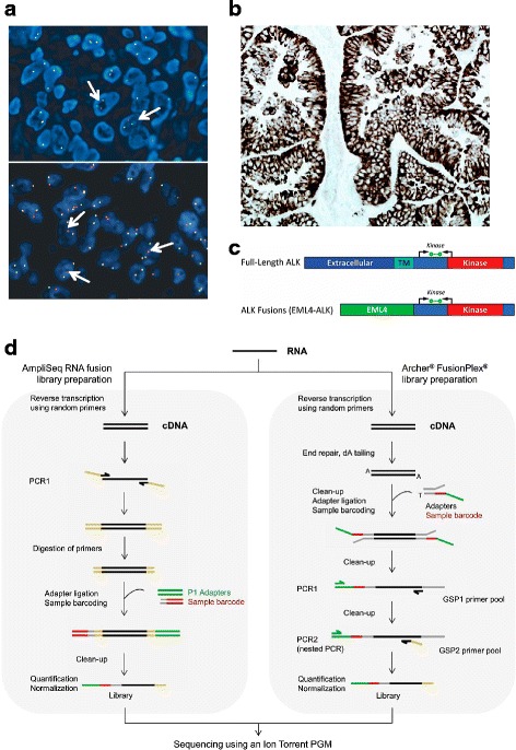
Fig. 2.

Diagnostic methods for the detection of ALK rearrangement and expression in NSCLC. a FISH: arrows in the upper picture exemplify the split signal pattern, while the ones in the bottom picture specified the single red signal pattern. b IHC using the D5F3 ALK assay. c Diagrammatic representation of full length ALK and the EML4-ALK fusion transcripts indicating ALK domains in the ALK protein, location of ALK RT-PCR primers (black arrows) and the fluorescent probe (green bar) used in the ALK RGQ RT-PCR Kit (Qiagen). TM: transmembrane. d Comparison of two commercially available methods to generate libraries for NGS. a and b adapted from ref. [45]. c reproduced from ref. [42]. d reproduced from ref. [46]
