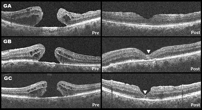
Fig. 2.

Representative cases from each group. The “pre” image is at baseline, the “post” image is at the 3 months follow-up. GA: Conventional 360 ILM peeling, displaying an asymmetrical U-shape closure configuration. In GB and GC (group B and C) the white arrowhead points to a hyperreflective area within the macular hole that may suggest retinal gliosis. The area is wider in the inverted-flap technique group. GB displays a symmetrical U-shape closure configuration. GC displays a symmetrical V-shape closure configuration
