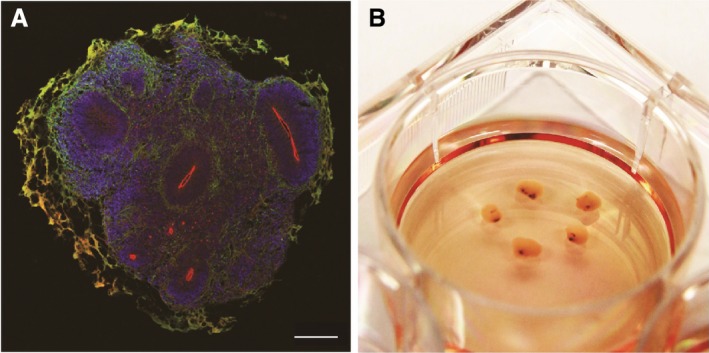
Figure 2.

A model of human midbrain organoids. (A) Developing (day 35) human midbrain organoids contained multiple polarized neuroepithelia. Apical side is marked by aPKC (red), basal side is enriched with neuronal cells marked by MAP2 (green). Note that the green‐orange color staining lining the exterior of the organoid is due to nonspecific staining of Matrigel. Scale bar: 500 um. (B) Long‐term cultures of human midbrain organoids accumulate neuromelanin (black spots).
