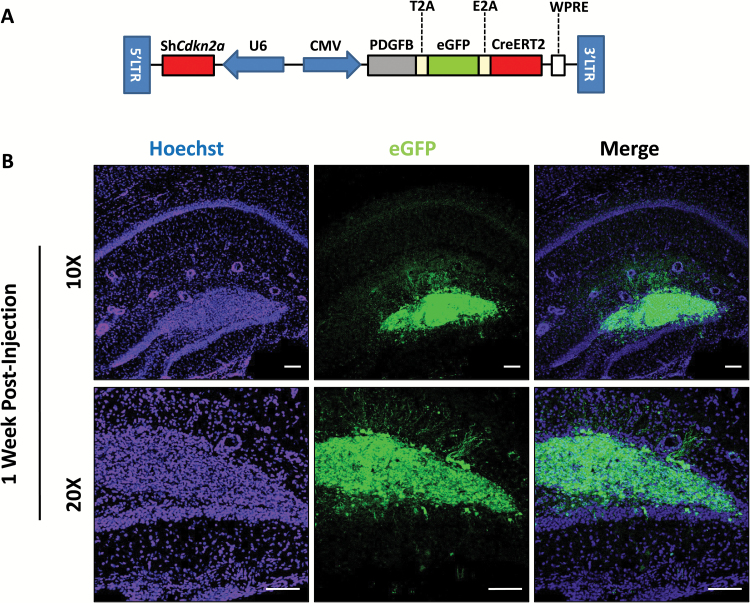
Fig. 4.
Fluorescent tracking of shCdkn2a-PDGFB-T2A-eGFP-E2A-CreERT2 transduced cells reveals early stages of tumorigenesis. (A) Vector design of shCdkn2a-PDGFB-T2A-eGFP-E2A-CreERT2 which expresses eGFP in the same coding locus as PDGFB and CreERT2. Infected cells in the dentate gyrus of an adult mouse (B) 1 week post lentiviral injection under 10X and 20X magnification. GFP (from the shCdkn2a-PDGFB-T2A-eGFP-E2A-CreERT2 lentivirus infected cells) and Hoechst (nuclei) channels are shown. Scale bar: 100 µm.

