Fig. 3.
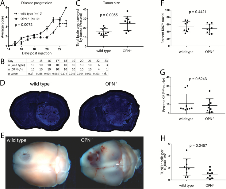
Loss of microenvironment-derived OPN expression enhances glioblastoma growth. (A) OPN−/− GL261-implanted animals show a significantly faster disease progression compared with wild type control animals (n = 10 animals per group). Disease scores are defined in Materials and Methods. Significance was calculated using linear regression analysis. (B) Animal numbers in each group and P-values showing significantly different disease scores between groups are listed for each day. (C) Tumors in OPN−/− mice are significantly larger than tumors in wild type control mice (n = 9 animals per group). (D) Representative 40-µm-thick sections of wild type and OPN−/− GL261-bearing mouse brains. Sections were stained with Hoechst. (E) Representative wild type and OPN−/− GL261-implanted brains 21 days post-injection. (F, G) No difference in the percentage of Ki67+ (F) and Ki67high (G) cells could be detected between tumors in OPN−/− and wild type mice. (H) Tumors in OPN−/− mice show less TUNEL+ cells per area than tumors in wild type mice. Analysis was done using Student’s t-test (B, C, F, G, H). Error bars represent SEM (A) or SD (C, F, G, H).

