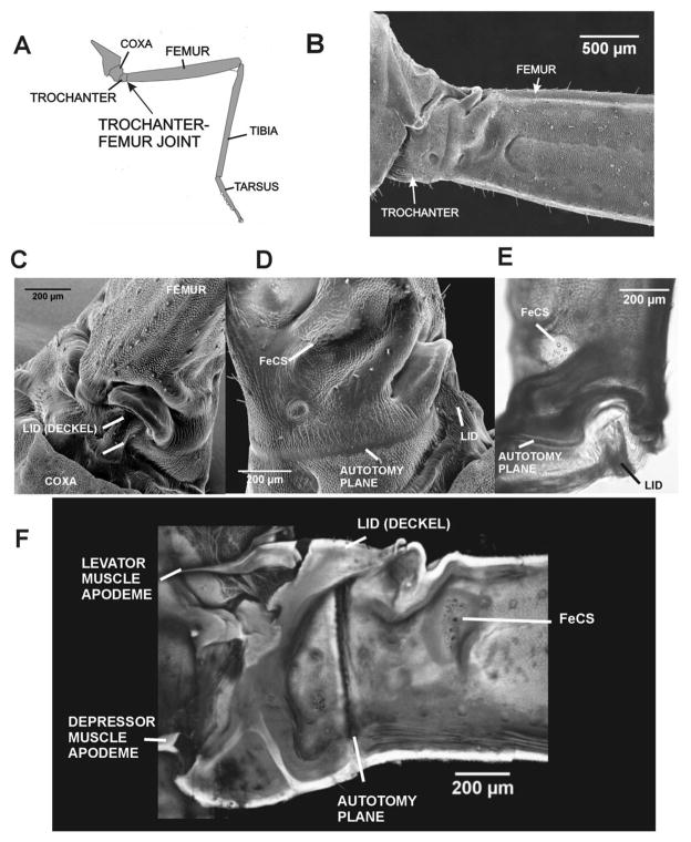
Fig. 2. Immobility and specializations of the trochanter-femur joint in stick insects.
A. Drawing of stick insect middle leg and trochanter-femur joint.. B. SEM of trochanter and femur in posterior view – The trochanter is functionally fused to the femur with no condyles or joint movement. C. SEM of upper (dorsal) surface of the trochanter – The dorsal surface of the trochanter forms a projection (termed the lid, G., Deckel) which interlocks with a depression on the proximal end of the femur. D. Posterior view (SEM) of proximal femur and lid – The femoral group of campaniform sensilla are located in a depression in the proximal femur, distal to the autotomy plane. E. Dorsal view of whole mount of TrF joint – the cuticle appears darkened and thickened in the region between the autotomy plane and femoral sensilla. F. Confocal projection image of inner (posterior) surface of a bisected coxa, trochanter and femur (the lid remained intact) – The apodeme (tendon) of the Levator Trochanteris muscle inserts upon the ‘lid’’ of the trochanter; the Depressor apodeme inserts upon the reinforced ventral end of the trochanter. The autotomy plane is clearly seen as a thin vertical line between the fused trochanter and femur.

