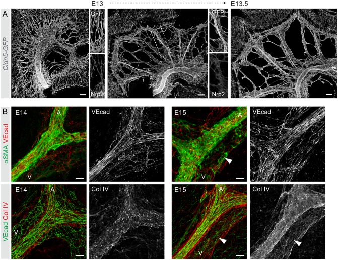
Fig. 2.
Mesenteric blood vasculature undergoes extensive remodeling and maturation between E13 and E15. (A) Visualization of the mesenteric vasculature in Cldn5-GFP embryos showing extensive remodeling from a primary plexus into a segmentally organized pattern of veins and arteries between E13 and E14. Single-channel images of the boxed areas show co-staining for the venous EC marker Nrp2 in only a subset of vessels. (B) Whole-mount immunofluorescence of E14 (left) and E15 (right) mesenteric vessels for markers of ECs (VE-cad; Cdh5), mural cells (αSMA; Acta2) and basement membrane (collagen IV). Single-channel images of indicated stainings are shown. Note poor EC alignment as well as mural cell and BM coverage in E14 vein (V) compared with E15 vein (arrowheads), or the artery (A). Scale bars: 100 μm (A); 20 μm (B).

