Fig. 3.
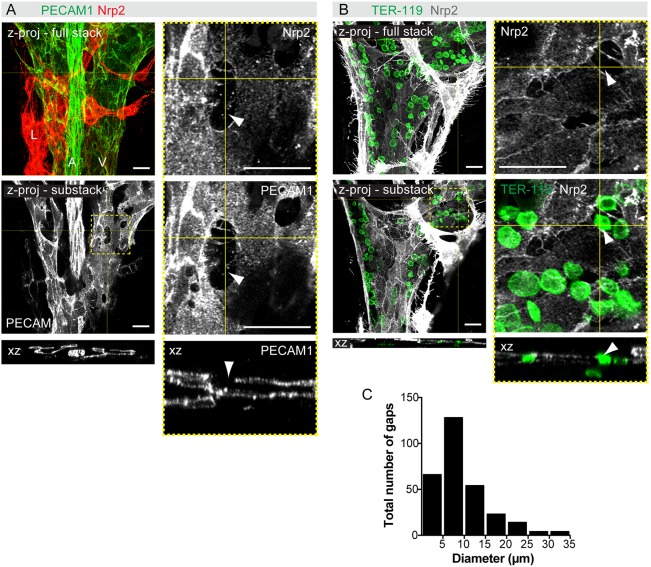
Remodeling of the mesenteric blood vasculature is associated with a transient loss of venous endothelial integrity. (A,B) Whole-mount staining of E14 mesenteries for the indicated antibodies, showing intercellular gaps in the veins. z-projections of confocal stacks are shown and boxed areas are magnified on the right. z-views at the indicated positions are shown below. Arrowheads indicate gaps in the endothelial layer. (C) Size distribution of intercellular gaps in wild-type E14 mesenteric veins (n=294 gaps from 14 embryos). Scale bars: 20 μm.

