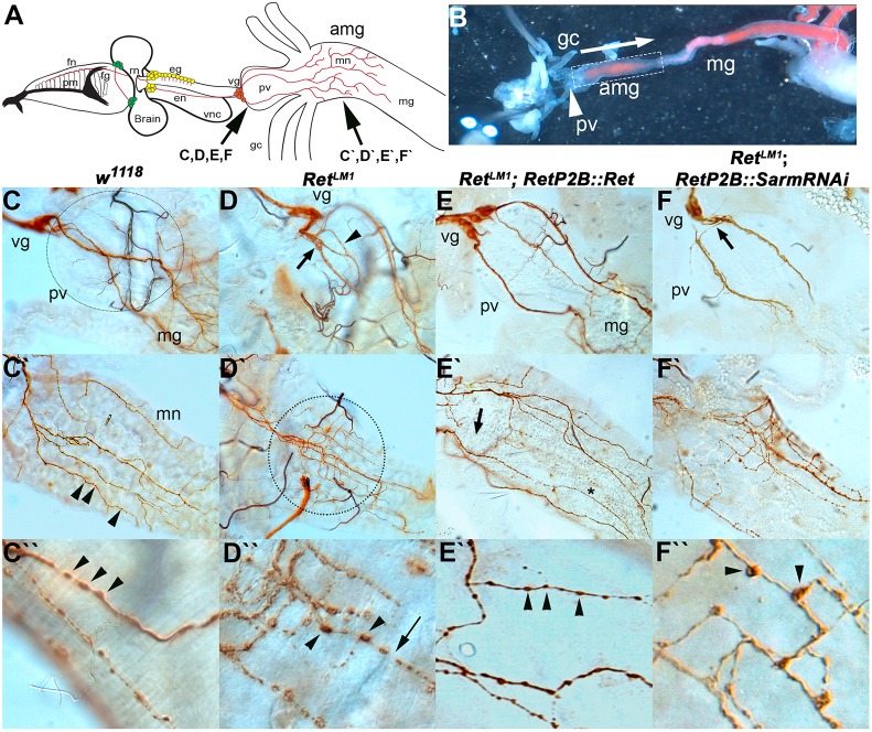
Fig. 4.
Larval midgut SNS architecture is dependent on Ret. SNS of second instar larvae was dissected and visualized with anti-Futsch (mAb 22c10) staining. (A) Larval SNS showing, from left, that the frontal nerve (fn) lies on top of the pharyngeal muscles (pm) and connects to the frontal ganglion (fg). The recurrent nerve (rn) projects anteriorly between the brain lobes to the esophageal ganglia (eg). The esophageal nerve (en) projects to the ventricular ganglion (vg) that lies at the anterior end of the proventriculus (pv). The midgut neuron (mn) axons project from the ventricular ganglion, over the proventriculus to innervate the anterior midgut (amg). The remainder of the midgut (mg) is not innervated. The ventral nerve cord (vnc) is also shown. (B) Dissected third instar larvae pinned in Drosophila S2 cell medium showing the relative positions of the proventriculus, anterior midgut and midgut. This preparation was used to observe spontaneous contractile activity in w1118 and RetLM1. Yeast paste dyed pink can be seen in the midgut. (C) In w1118 animals, the proventriculus (unstained, dotted circle indicates extent) forms at the junction of the foregut and midgut and acts as a valve regulating entry of food into the midgut. The proventriculus and midgut are innervated by cells of the ventricular ganglion, which are located on the anteriormost point of the proventriculus. Axons of the midgut neurons (brown) traverse the proventriculus as two or three tightly fasciculated bundles and only start branching on reaching the midgut. (C′) On encountering the midgut, the midgut neuron axons (brown) branch in a regular fashion to spread out over the anterior end of the midgut. (C″) Multiple small varicosities can be seen throughout the midgut axons as punctate increases in 22c10 staining (C′,C″, arrowheads). (D) RetLM1 allele with midgut SNS defects typically seen in Ret mutants. The midgut nerve axons defasciculate (arrow) a very short distance from the ventricular ganglion and show additional defasciculation (arrowhead) as they traverse the proventriculus. (D′) Upon encountering the midgut, they fail to spread out and remain confined to a much smaller area than in w1118 (dotted circle). Within this area, the midgut neuron axons display a much higher level of branching. (D″) Axonal varicosities appear larger and the distribution of the 22c10 antigen is reduced between varicosities. The appearance initially suggested that axon fragmentation is occurring. (E) Rescue of Ret mutant phenotypes using UAS-Ret driven by RetP2B-GAL4 in a RetLM1 genetic background. Axons from the ventricular ganglion crossing the proventriculus more closely resemble the axon bundles in w1118, remaining tightly fasciculated until the midgut is reached. (E′) Midgut of RetLM1 RetP2B-GAL4::UAS-Ret larva in which the axon pattern resembles w1118, with the axons spreading out to cover a large proportion of the anterior midgut. The branching frequency also resembles that in w1118. (E″) Axonal varicosities resemble w1118, being smaller with a more even distribution of 22c10 antigen between the varicosities. (F) RetP2B-GAL4 driving UAS-SarmRNAi does not rescue the aberrant branching caused by RetLM1, as the proventriculus nerve bundles defasciculate rapidly after leaving the cell bodies of the ventricular ganglion (arrow) and continue to show defasciculation as the axons traverse the proventriculus. (F′) The mn axons also branch more frequently and do not cover the same area of midgut as in w1118. (F″) The increased prominence of axonal varicosities observed in Ret mutants remains.

