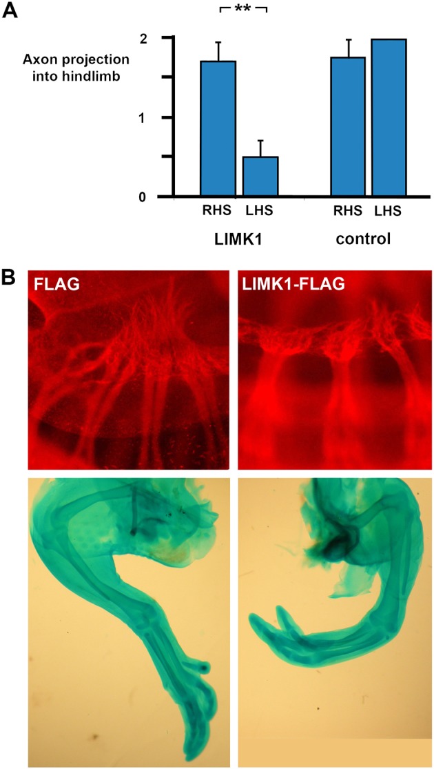
Fig. 10.

Electroporation of LIMK1 into chicken neural tube causes axon loss and clubfoot. Electroporation of plasmids expressing LIMK1 or empty vector controls into HH stage 11 chickens followed by immunohistochemistry for β-III-tubulin 72 h later or Alcian Blue cartilage staining after a further 5 days. (A) Nerve projection scored 0-2 as described in the Materials and Methods for electroporated (left-hand side, LHS) and non-electroporated contralateral sides (right-hand side, RHS) of each embryo, for LIMK1 and control vectors, and shows significant inhibition of axon growth in LIMK1-treated nerves, but not in empty-vector electroporations. LIMK1 electroporation: RHS nerve score=1.67±0.25, n=9; LHS score=0.5±0.22, n=8 (one embryo damaged cf. RHS). Control FLAG electroporation: RHS nerve score=1.75±0.25, n=4; LHS score=2±0.00, n=3. **P=0.001 (paired t-test). Error bars represent s.e.m. (B) (Top) Whole-mount β-III-tubulin immunohistochemistry (red) on chicken embryos showing normal sciatic plexus formation and axon projection (score 2) after an empty ‘FLAG’ vector transfection compared with representative failure of nerve plexus formation (score 0) after LIMK1 transfection. (Bottom) Alcian Blue cartilage preparation of (left) a control FLAG-electroporated chicken limb and (right) limb of one of the chickens (3/12) that exhibited a mild clubfoot-like phenotype after transfection with LIMK1.
