Fig. 4.

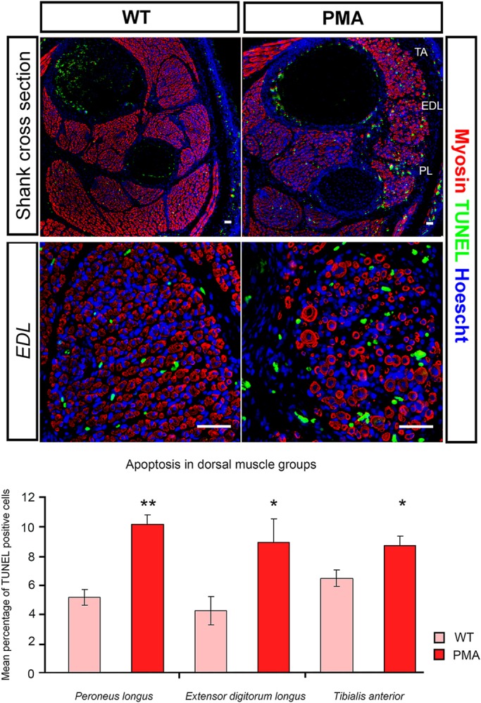
Increased apoptosis in dorsal muscle blocks of the pma hindlimb. Top: TUNEL labelling (green) to visualise apoptotic cells in cross-sections of E16.5 wild-type (WT; left) and pma/pma foetuses (right), combined with immunohistochemistry for myosin heavy chain (red) and Hoechst nuclear stain (blue). Higher magnification of the one dorsal muscle, the extensor digitorum longus, is shown. Bottom: Although apoptosis occurs in all muscles, the percentage of TUNEL-positive cells was significantly greater in the three major dorsal muscle blocks of pma/pma foetuses than in wild-type controls (n=8 for both groups). *P<0.05; **P<0.01. Error bars represent s.e.m. Scale bars: 50 µm.
