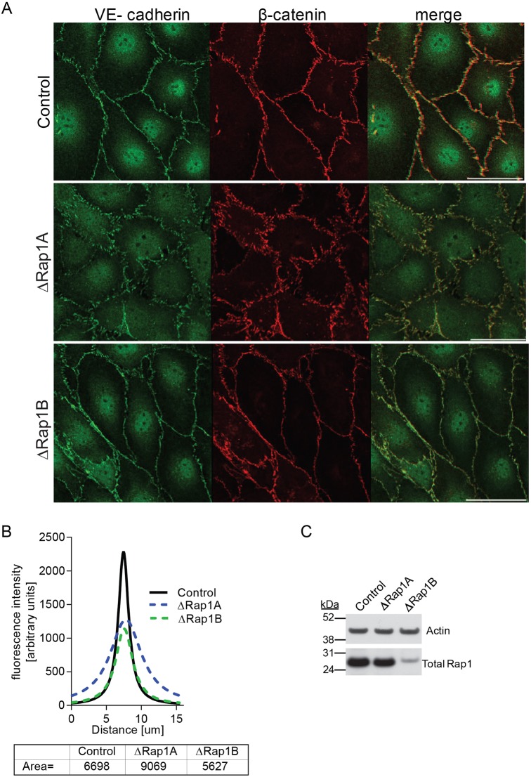
Fig. 2.
Rap1A deficiency leads to disorganized AJs. (A) Confocal images of control, ΔRap1A or ΔRap1B primary mouse lung ECs (MLECs) grown to confluence and stained for AJ proteins, as indicated. Dispersed VE-cadherin and β-catenin staining is visible in ΔRap1A ECs, but not in ΔRap1B ECs. The same β-catenin-stained control and ΔRap1A MLECs are also shown in Fig. 5A. Scale bar: 20 μm. (B) Fluorescence intensity distribution across cell–cell junctions was measured in three independent images with 10 randomly selected contacts per experimental condition. After correcting for background fluorescence, data were nonlinear Lorentzian curve-fitted to each profile using Prism5, as previously described (Smutny et al., 2010) and area under the curve was calculated (bottom). (C) Representative immunoblots of total Rap1 content in Rap1-deficient ECs; actin is shown as loading control. Control and ΔRap1B MLECs were isolated from control (Tie2-Cre0/0; rap1a+/+rap1bf/f) or Rap1B-ECKO (Tie2 Cre+/0; rap1a+/+rap1bf/f) P6-9 lungs. ΔRap1A ECs were obtained by knocking down Rap1A in control cells using ‘pooled’ siRap1A.

