Fig. 5.
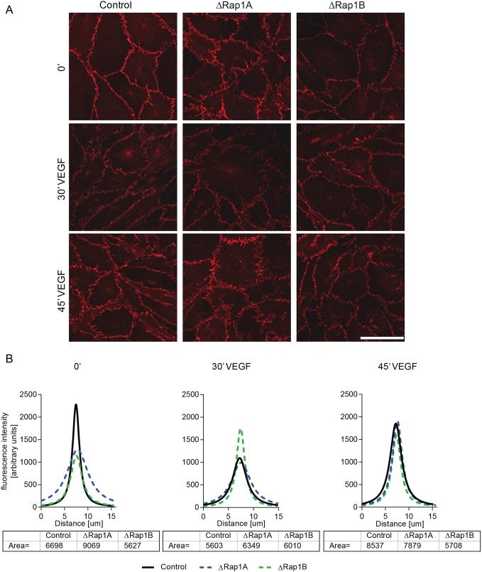
Rap1b deficiency impairs the ability of VEGF to remodel AJs. (A) Confocal images of β-catenin staining in ΔRap1A, ΔRap1B or control confluent MLECs, serum-starved for 6 h as in Fig. 2 (control and ΔRap1A MLECs are reproduced from Fig. 2A) and treated for indicated time with 50 ng/ml VEGF. Scale bar: 20 μm. (B) AJ fluorescence intensity distribution and calculation of area under the curve at corresponding time points of VEGF treatment, measured as described in Fig. 2. After 30 min of VEGF treatment, AJs are dispersed in WT and ΔRap1A ECs, but not in ΔRap1B ECs (A, middle row), in which fluorescence intensity is unchanged (B, middle graph). Representative images of n=3 independent experiments are shown.

