Fig. 1.
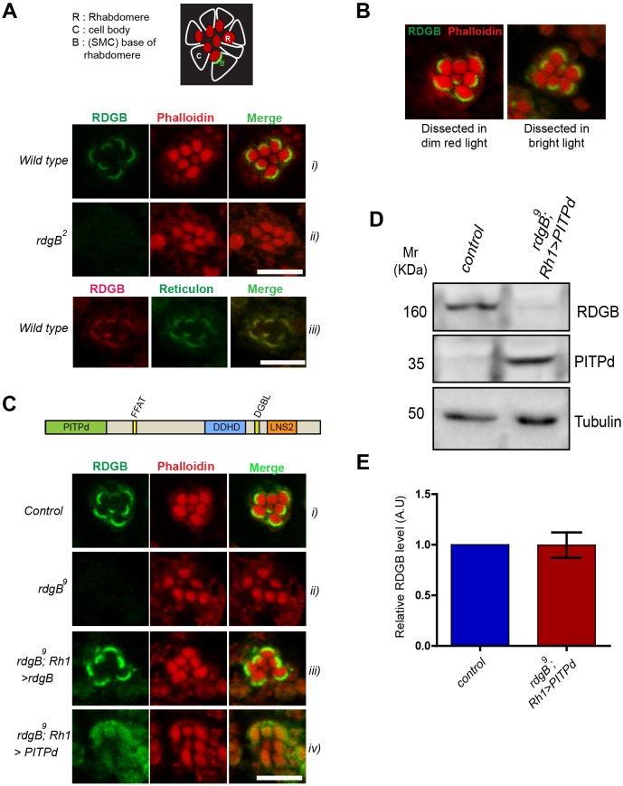
The PITP domain of RDGBα could not rescue all functions of RDGBα. (A) Localization of endogenous RDGBα in PRs using antibody against the PITP domain. (i) Confocal images showing localization of RDGB in wild-type PRs of 1-day-old flies. Phalloidin stains actin and marks the rhabdomeres. (ii) rdgB2 (null) shows no staining. (iii) RDGBα co-localizes with the reticulon as detected by anti-GFP staining of a reticulon::GFP fusion protein. Top panel is a cartoon representation of an ommatidium showing arrangement of PRs and the base of rhabdomeres. (B) Confocal images showing localization of RDGBα in wild-type PRs when dissected in dim red light versus bright light. (C) Localization of exogenously expressed RDGB and its PITPd. Confocal images showing localization of RDGBα in 1-day-old flies expressing rdgB and PITPd using Rh1Gal4 promoter. (D) Representative western blot image showing level of expression of Rh1>PITPd in rdgB9 head extracts. (E) Quantification of band intensity depicted in D. Error bars represent s.e.m. (n=4 blots). Scale bars: 5 µm.

