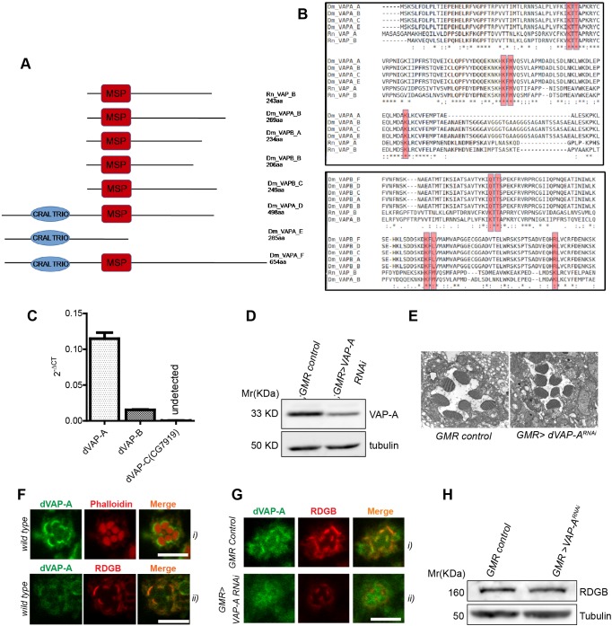
Fig. 3.
dVAP-A is enriched at the SMC and is essential for RDGBα localization. (A) Protein structure of Drosophila VAP proteins (dVAP-A and dVAP-B) and R. norvegicus VAP proteins (Rn VAP_A and Rn VAP_B). The structure of alternative splice variants of dVAP-A and dVAP-B are shown, as are the major sperm protein (MSP) domain and the CRAIL-TRIO domain. (B) Multiple sequence alignment of the Drosophila and mammalian VAP protein isoforms. Asterisk indicates conservation, dot marks lack of conservation. The conserved residues that coordinate with the FFAT motif of interacting proteins is shown within the red boxes. See Materials and Methods for protein ID. (C) Expression levels of dVAP mRNA in wild-type Drosophila retinae. RP49 was used as housekeeping gene and transcript levels for dVAP isoforms are shown normalized to RP49. (D) Representative immunoblot and quantification showing expression level of Dm-VAP-A in retinae extract of flies of mentioned genotypes (n=2 blots). (E) Representative scanning electron micrograph showing a single ommatidium of 1-day-old flies of indicated genotypes. (F) Confocal images showing localization of endogenously expressed dVAP-A in photoreceptors (i) and co-localization with RDGB (ii). (G) Confocal images showing mis-localization of RDGB in GMR>VAP-ARNAi. (H) Representative immunoblot showing expression level of RDGB in GMR>VAP-ARNAi compared with GMRGal4/+ flies. Scale bar: 5 µm.

