Fig. 4.
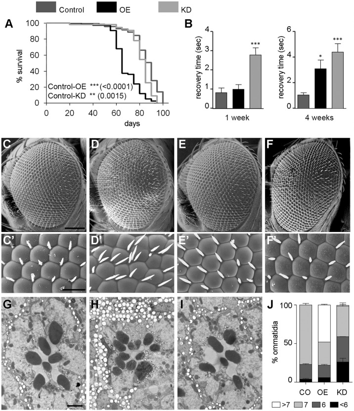
Neuronal alterations in the OE and KD genotypes. (A) Survival curves of the control (elav-Gal4), OE and KD flies; log-rank (Mantel–Cox) test shows significant differences with the control genotype. (B) Bang-sensitivity analyses of the three genotypes at 1 and 4 weeks of age, represented as recovery time after mechanical stress-induced paralysis. (n=4, ≥10 flies per experiment). (C,C′) SEM image of a wild-type control eye (GMR-Gal4, C) and higher magnification showing the stereotypical hexagonal arrangement of the ommatidia and the inter-ommatidial bristles (C′). (D,D′) In an OE fly, the eye has a rough aspect (D), and the ommatidia have lost the regular pattern and have supernumerary bristles (D′). (E,E′) KD fly eyes have a wild-type aspect (E) and structural arrangement (E′). (F,F′) The defects in UAS-jp eyes are corrected by coexpression of UAS-jpRNAi. (G) SEM of an ultra-thin section of a control fly ommatidium, showing the wild-type trapezoidal arrangement with seven rhabdomeres. (H) Structure of an ommatidial section of an OE fly with extra rhabdomeres, as a result of the recruitment of extra photoreceptor neurons. (I) Loss of rhabdomeres, hence photoreceptor neurons, in a KD ommatidial section. (J) Distribution of the number photoreceptor neurons in ommatidia of the three genotypes at 1 week of age (n=4, ≥60 ommatidia per section). Scale bars: 100 μm in C-F; 20 μm in C′-F′; 2 μm in G-I. In bar diagrams, data are represented as mean±s.e.m. One-way ANOVA, *P<0.05.

