
Fig. 2.
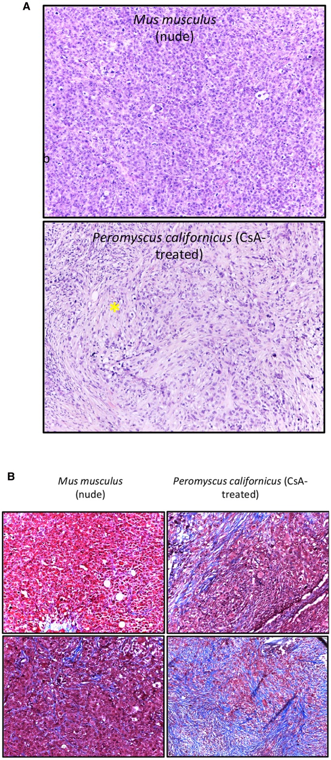
Histology of MDA-MB-231 tumors. (A) H&E-stained sections of tumors from MDA-MB-231 human breast cancer cells admixed with human HFFF2 fibroblasts in nude mice (upper panel) or P. californicus (lower panel). The asterisk indicates areas with spindling cells. Magnification, 10×. (B) Trichrome-stained sections of tumors from MDA-MB-231 human breast cancer cells admixed with human HFFF2 fibroblasts in nude mice (left) or P. californicus (right). Blue staining indicates the presence of collagen. Images from two tumors from each group are shown, selected to contain the lowest (top) and the highest (bottom) amounts of collagen. Magnification, 20×.