Fig. 3.
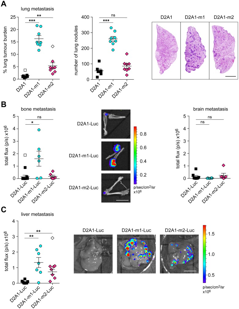
Experimental metastasis assays. (A) 4×10⁵ D2A1, D2A1-m1 or D2A1-m2 cells were inoculated via the tail vein into BALB/c mice (n=8 mice per group). 11 days later, lungs were removed at necropsy. Data show quantification of tumour burden as monitored by % tumour area and number of lung nodules per lung section. Representative lung images are shown (right). Scale bar: 2 mm. (B) 2×10⁵ D2A1-Luc, D2A1-m1-Luc or D2A1-m2-Luc cells were inoculated into the left ventricle of BALB/c mice (n=6 mice per group). 10 days later, bones and brains were removed at necropsy and IVIS imaged ex vivo. Representative ex vivo bone IVIS images are shown (middle). Scale bar: 1 cm. (C) 2×10⁵ D2A1-Luc, D2A1-m1-Luc or D2A1-m2-Luc cells were inoculated into the spleen of BALB/c mice (n=8 mice per group). 13 days later, livers were removed at necropsy and IVIS imaged ex vivo. Representative IVIS images are shown (right). Scale bar: 1 cm. Significant outliers are shown as white symbols. All data are mean values per mouse ±s.e.m. *P<0.05; **P<0.01; ***P<0.001; ns, nonsignificant.

