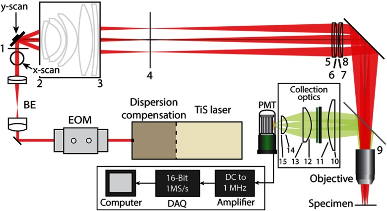
Fig. 7.
LF-TPM system schematic. Pulsed light from TiSapphire (TiS) laser is directed to the input of the microscope. Laser intensity and dispersion are controlled with an electro-optic modulator (EOM) and dispersion compension prisms. The beam is then expanded with a beam expander (BE) consisting of two achromatic doublets. Emission is separated with a dichroic mirror and is transmitted through both a shortpass and notch filter before being collected by a photomultiplier tube (PMT). The output of the PMT is amplified and digitized before images are displayed on a computer. Surfaces are labeled with numbers, and the distances are presented in Appendix C.

