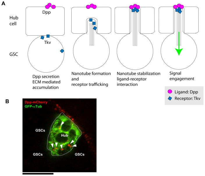
Figure 2. MT-nanotube mediated niche-stem cell signaling.
(A) Model for MT-nanotube mediated signaling. Dpp induces MT-nanotube formation, and receptor–ligand interaction occurs at the surface of MT-nanotubes, leading to signaling activation in GSCs. (B) Dpp-mCherry (red) expressed in hub cells together with GFP-α-Tubulin (green, hub cell cortex) using hub specific unpaired (Upd) promoter. DppmCherry forms punctae along hub cell cortex (arrowheads). Scale bar: 10μm. Entire hub area is encircled by white broken line. GSCs are attached to hub from surrounding area (not visible here).

