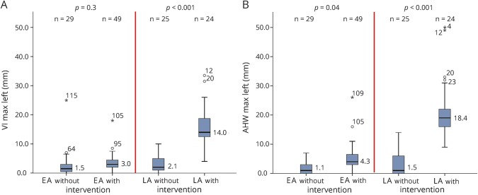
Figure 2. Median maximum ventricular measurements for infants without and with intervention in EA and LA groups.
Maximum ventricular measurements, including VI in millimeters above the +2 SD line adjusted for postmenstrual age on the y-axis in panel A and AHW in millimeters above the 6 mm line on the y-axis in panel B, for the infants without and with intervention in the EA and the LA groups depicted on the x-axis. Similar graphs were obtained for median maximum VI and AHW measurements for the right lateral ventricles. AHW = anterior horn width; VI = ventricular index.

