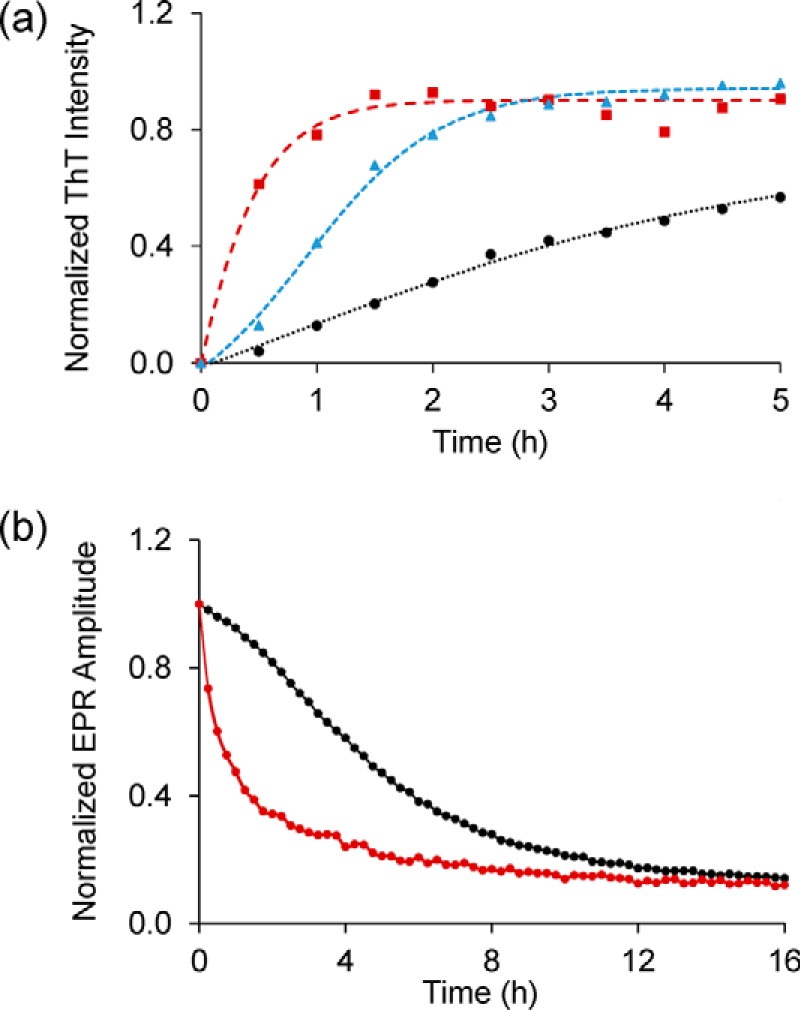
Figure 4.

Membranes strongly enhance aggregation of Httex1(Q46). a, ThT fluorescence kinetics for 35R1 Httex1(Q46) derivative in the presence (red) or absence (black) of vesicles (25% POPS/75% POPC, 375 μm total lipid concentration). Also shown for comparison are the seeded kinetics from Fig. 3 (blue). b, membrane-mediated enhancement in aggregation can also be detected by EPR using the 35R1 derivative of Httex1(Q46). The control without membranes is shown in black, whereas the EPR data obtained in the presence of 25% POPS/75% POPC vesicles (375 μm) are in red. The protein concentration was 15 μm in all cases.
