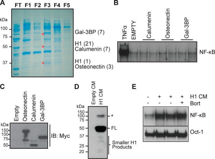
Figure 1.
Identification of HAPLN1 as a factor capable of inducing NF-κB activity in myeloma cells. A, SDS-PAGE and Coomassie Blue staining of the enriched fractions with NF-κB–inducing activity previously identified by Markovina et al. (35). Three bands (*) from fraction 3 (F3) that contained the highest NF-κB–inducing activity were excised and analyzed by nano-LC-MS/MS. Shown are identified factors with the number of unique peptides identified in parentheses. B, EMSA analysis of RPMI8226 cells incubated with 10 ng/ml TNFα for 15 min or CM collected from HEK293 transiently transfected with vector containing C-terminally MYC-tagged proteins: calumenin, osteonectin, or galectin-3–binding protein (Gal-3BP) for 2 h. Results are representative of at least three independent experiments. C, representative immunoblot analysis (IB), using anti-MYC antibody, of CM from HEK293 cells transiently transfected with calumenin, osteonectin, or galectin-3–binding protein expression vector showing the presence of corresponding secreted factors. D, immunoblot analysis, using anti-MYC antibody, of HAPLN1 expression in CM collected from transiently transfected HEK293 cells with the HAPLN1 expression vector positive for induction of NF-κB activity. Smaller HAPLN1 (H1) fragments are indicated *, larger immunoreactive bands. E, EMSA analysis of RPMI8226 cells treated with CM from transiently transfected HEK293 cells containing HAPLN1 for 2 h in the absence or presence of 100 nm bortezomib (Bort). Induced NF-κB activity is highly variable in more than five independent experiments, possibly dependent on the variability of smaller H1 species seen in D.

