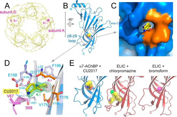
Figure 3.
X-ray crystal structure of α7-AChBPVS in complex with allosteric binder fragment CU2017. A, yellow ribbon representation of the α7-AChBPVS pentamer as seen along the 5-fold symmetry axis with the bottom (C-terminal side) of the pentamer pointing toward the viewer. The magenta mesh is the anomalous difference density at a contour level of 4σ. B, schematic representation of the A subunit with fragment CU2017 bound to the β8–β9 loop site. Fragment CU2017 is shown in transparent ball & stick representation in yellow (carbon), blue (nitrogen), red (oxygen), and magenta (bromine). C and D, detailed surface and ball and stick representation of the β8–β9 loop site. Residues belonging to the human α7 nAChR are colored in blue and residues from Lymnaea AChBP are colored in orange. Residues Val-67 and Ser-68 (pink) are from a neighboring pentamer in the crystal packing. In D, the green mesh is the simple difference density at a contour level of 4σ, the magenta mesh is the anomalous difference density at 4σ. E, comparison of the β8–β9 loop site occupied by fragment CU2017 and previously published structures of the Erwinia ligand-gated ion channel ELIC in complex with chlorpromazine (28) (PDB code 5lg3) or bromoform (27) (PDB code 3zkr).

