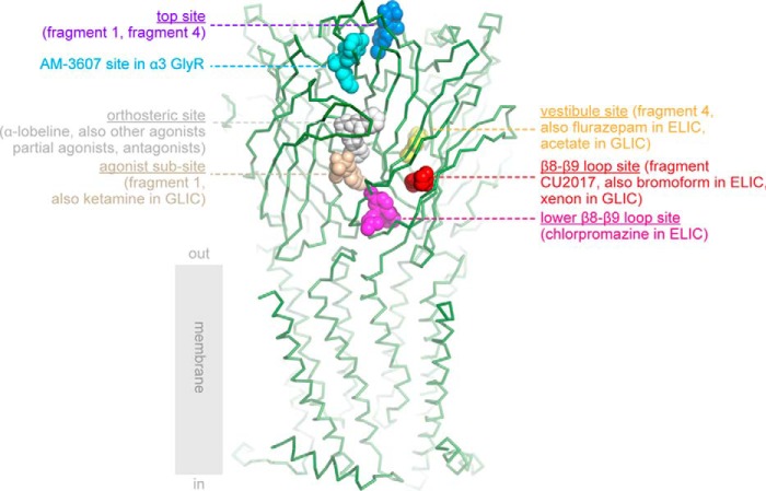
Figure 5.
Overview of ligand-binding sites in α7-AChBP and ligand-binding domains of other Cys-loop receptors. The crystal structure of α7-AChBPVS is shown in green ribbon representation and was superposed onto the extracellular domain of the human α4β2 nAChR structure (13) (PDB code 5kxi). Only the transmembrane domain of the α4β2 receptor is shown for illustrative purposes. The different binding sites are indicated according to their respective bound ligands in different Cys-loop receptor structures and corresponding PDB accession codes are mentioned between parentheses: top site in blue, α7-AChBP + fragment 1 (17) (code 5afj) and α7-AChBPVS + fragment 4 (this study) (code 5oug); AM-3607 site in cyan, α3 GlyR + AM-3607 (43) (code 5tin); orthosteric site in white, example α7-AChBPVS + α-lobeline (code 5ouh); agonist subsite in wheat, α7-AChBP + fragment 1 (17) (code 5afj) and GLIC + ketamine (49) (code 4f8h); β8–β9 loop site in red, α7-AChBPVS + fragment CU2017 (this study) (code 5oui), ELIC + bromoform (27) (code 3zkr) and locally closed GLIC + xenon (47) (code 4zzb); lower part of the β8–β9 loop site in magenta, ELIC + chlorpromazine (28) (code 5lg3); vestibule site in yellow, AChBPVS + fragment 4 structure (this study) (code 5oug), ELIC + flurazepam (18) (code 2yoe), and GLIC + bromoacetate (44) (code 4qh1).

