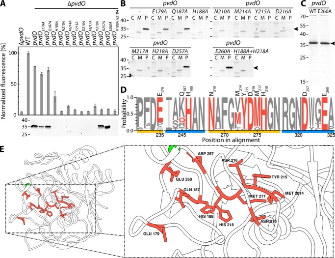
Figure 7.
Effects of alanine exchanges of residues with potential catalytic function. A, effect of the indicated single-alanine exchanges on the formation of fluorescent pyoverdine and control of the presence of the respective PvdO variants in the periplasm by SDS-PAGE/Western blotting. B, subcellular fractionation of all analyzed PvdO variants. Cytoplasmic (C), membrane (M), and periplasmic (P) fractions were analyzed for the presence of PvdO by SDS-PAGE/Western blotting. Note than in case of stable PvdO variants, the protein is exclusively detectable in the periplasm, as expected for Sec transport. C, Coomassie-stained PvdO preparations that had been used for metal detections. D, sequence logo of the mutated regions (see “Experimental procedures” for details). Exchanged residues in PvdOA506 are colored red, and distinct regions are highlighted in yellow and blue. The residue numbers on top of the sequence logo indicate the residue position in PvdOA506. E, homology model of the PvdOA506 structure based on the known structure of PvdO from P. aeruginosa (PDB code 5HHA). Exchanged residues are highlighted in red. Left, overview of the structure; right, close-up view. The model was visualized using Chimera (36). Masses of marker proteins (in kDa) are indicated on the left side of blots.

