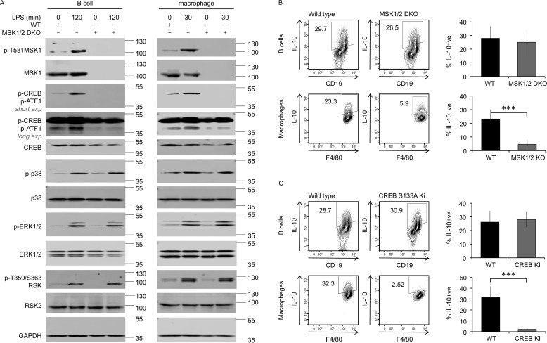
Figure 4.
MSK and CREB regulate LPS–induced IL-10 production in macrophages but not B cells. A, B-cell and macrophage populations were isolated from peritoneal cavity cells from wildtype and MSK1/2 double knockout mice. The cells were treated for the indicated times with 10 μg/ml LPS or left unstimulated. The levels of the indicated proteins were assessed in cell lysates by immunoblotting. Long and short exposures of the phosphor-CREB/ATF1 blot are shown to allow visualization of both phospho-CREB (upper band) and phospho-ATF1 (lower band). B and C, peritoneal cavity cells from wildtype, MSK1/2 double knockout (B), and CREB S133A knockin (C) mice were stimulated with 10 μg/ml LPS + brefeldin A + monensin for 5 h. Intracellular IL-10 in B cells and macrophages was assessed by flow cytometry. Representative plots are shown in the left panels. The data shown in the right panels represent the averages and standard deviations of biological replicates (n = 12 in B, n = 6 in C). ***, p < 0.001 (two-tailed unpaired Student's t test).

