Figure 1.
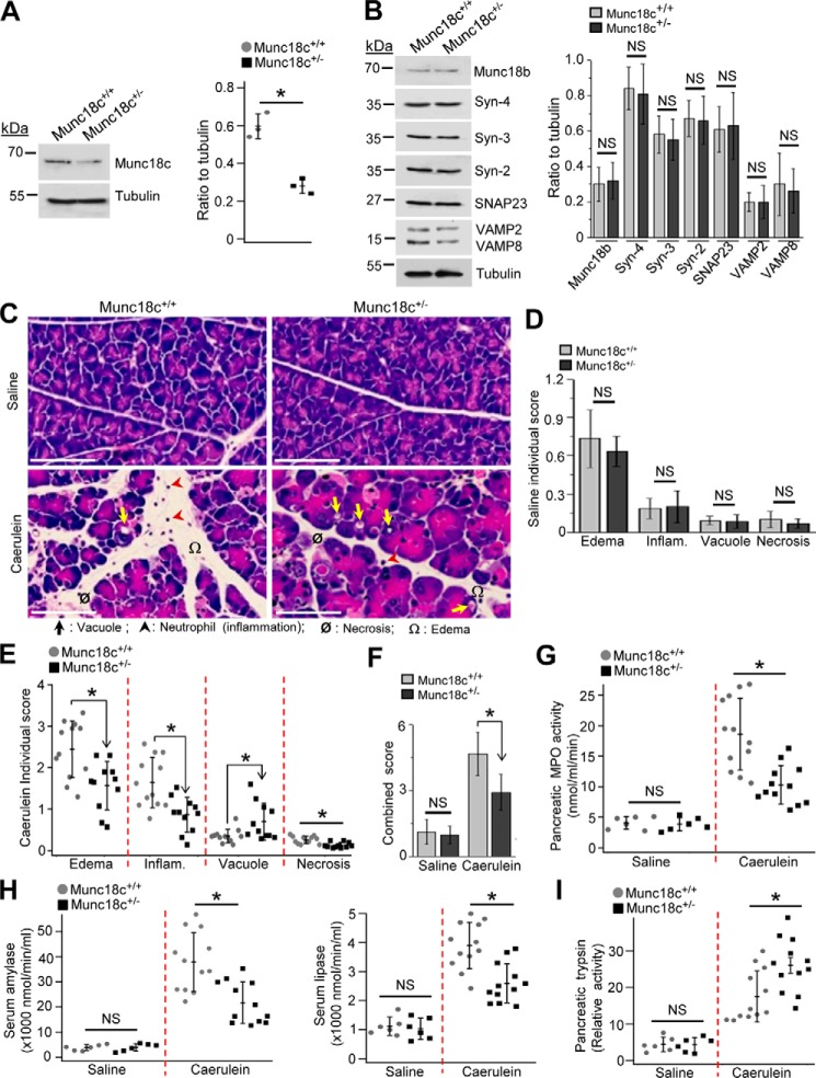
Munc18c depletion in mice protects against caerulein-induced pancreatitis. A, representative Western blots (top panel, n = 3) and densitometry analysis (right, normalized to tubulin) showing 52% reduced expression of Munc18c in Munc18c+/− pancreatic acini. B, representative Western blots (left panel, n = 3) showing no change in expression (right panel, densitometry normalized to tubulin) of indicated proteins in WT and Munc18c+/− mouse acini. C, representative H&E-stained images of pancreas from control (top panels, 8 hourly intraperitoneal injections of 0.9% saline) and caerulein-administered (bottom panels, 8 hourly intraperitoneal injections of 50 μg/kg) WT (left) and Munc18c+/− (right) mice. Scale bars, 50 μm. Edema, inflammation, vacuolization, and necrosis are indicated by symbols. Larger area of histology images with magnified regions are in Fig. S1, A and B to better display the pancreatic injury. D and E, individual histology scores (on a scale of 0–4) of control (D, n = 6) and caerulein-treated (E, n = 12) pancreas. F, combined histology scores from D and E. G and H, quantitative measurements of activities of pancreatic myeloperoxidase (MPO) (G), serum amylase (H, left) and serum lipase (H, right) in control or caerulein-administered WT and Munc18c+/− mice. I, relative activity of pancreatic trypsin in control or caerulein-administered WT and Munc18c+/− mice. Enzyme activities were assayed on tissue samples from mice that were used for analysis in (D–F). Data with nonsignificant differences are expressed as ± S.D. Data with statistically significant differences are shown as scattered plots with S.D. *, p < 0.05, NS, not significant.

