Figure 6.
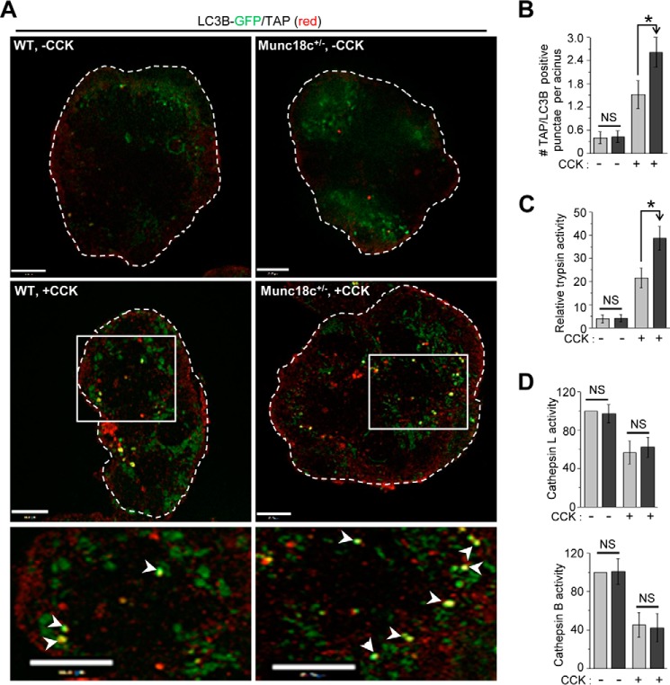
Munc18c depletion promotes autolysosomal trypsinogen activation. A, representative merged confocal immunofluorescence images of LC3B GFP (green) and TAP (red) in WT and Munc18c+/− mouse acini that were either kept as control (no CCK-8, top panels) or stimulated with 10 nm CCK-8 for 30 min (bottom panels). Selected areas from the stimulated images are magnified to better view their co-localization shown as yellow hotspots (pointed to with white arrowheads) indicative of activated trypsin within the autolysosomes. n = 3 independent experiments. Scale bars, 10 μm. Full sequence of images are shown in Fig. S5. B, quantification of TAP-positive LC3B GFP puncta from 40 acinar cells from three independent experiments performed as described previously (7). Data expressed as mean ± S.D. C, relative activity of trypsin in lysates from WT and Munc18c+/− mouse acini stimulated as in (A). Data expressed as mean ± S.D. from three independent experiments. D, relative activities of cathepsin L (top) and cathepsin B (bottom) in the lysosomal fractions from WT and Munc18c+/− mouse acini that were either kept as control or CCK-stimulated as in (A). The activities of cathepsins in controls were taken as 100. Data expressed as mean ± S.D. from three independent experiments. *, p < 0.05, NS = not significant.

