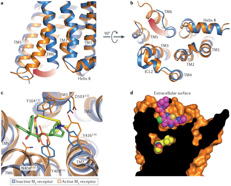
Figure 4. Activation and allosteric modulation of the M2 receptor.

As shown in part a and part b, the intracellular tip of transmembrane domain 6 (TM6) rotates outwards in the active M2 receptor structure (orange) relative to the inactive state (blue). As shown in part c, the orthosteric binding site contracts upon M2 receptor activation, enclosing the agonist iperoxo (yellow) in a smaller binding site, as compared to the antagonist (QNB; green) binding cavity. Residues are numbered according to the human M2 receptor sequence. As shown in part d, LY2119620 (magenta), a muscarinic positive allosteric modulator, binds to the extracellular vestibule of the M2 receptor directly above the orthosteric agonist iperoxo (yellow). ICL2, intracellular loop 2.
