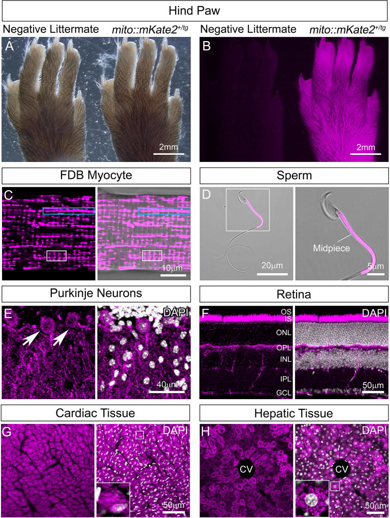
Figure 2. Widespread mitochondrial labeling in the CAG-mito::mKate2 transgenic line.
Hind paw of a CAG-mito::mKate2+ transgenic founder progeny showing bright mKate2 fluorescence as compared to a negative littermate (A–B). Expression of mito::mKate2 and resolution of tissue-specific mitochondrial morphologies and distribution in the adult flexor digitorum brevis (FDB) muscle (C), sperm midpiece (D), Purkinje neurons of the cerebellum (E), retina (F), heart (G) and liver (H). GCL=Ganglion Cell Layer, IPL=Inner Plexiform Layer, INL=Inner Nuclear Layer, OPL=Outer Plexiform Layer, ONL=Outer Nuclear Layer, IS=Inner Segments, OS=Outer Segments, CV=Central Vein. White nuclear stain is DAPI. n = 3.

