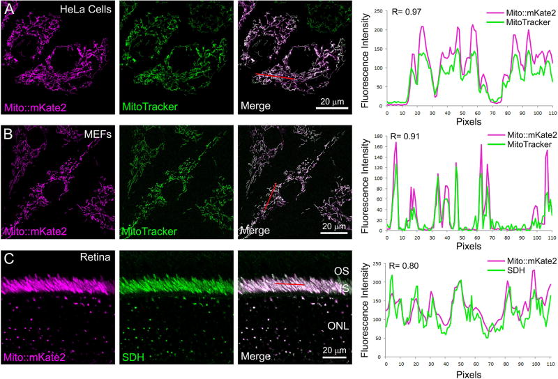
Figure 3. Co-localization of mito::mKate2 fluorescence and mitochondrial-specific markers.
HeLa cells transfected with the CAG-mito::mKate2 plasmid and labeled with MitoTracker Green (A). Mito::mKate2 transgenic MEFs co-labeled with MitoTracker Green (B). Mito::mKate2 transgenic retinal cryosections stained with anti-SDH antibodies (C). Red lines demarcate pixels used for co-localization analyses. n = 3. See text for details.

