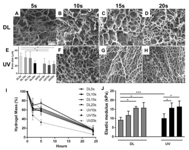
Figure 6.
Morphology (DL images A to D, UV images F to H) and pore size (E), degradation (I) and elastic modulus (J) of GelMA hydrogels. SEM images showed microporous structure induced during polymerization of GelMA hydrogels. The porous structure of DL photopolymerized hydrogels at different time points are larger than UV photopolymerized hydrogels. UV photopolymerized GelMA hydrogels degraded faster than DL photopolymerized GelMA hydrogels after 10s exposure. The elastic modulus increased with the exposure time for both DL and UV photopolymerized GelMA hydrogels. p<0.05 (*).

