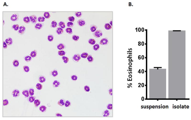
Figure 2. Evaluation of live CD11c-Gr1-/loMHCII- eosinophils by light microscopy.

A. Light microscopic image of a larger field of eosinophils isolated from lungs of IL5tg mice by the FACS protocol as shown in Fig. 1. Image photographed with a Leica DMI4000 microscope with camera; photographs taken at 40X magnification. B. Percent eosinophils in the initial single cell suspension and in the post-FACS isolate; > 200 cells counted per sample.
