Figure 4.
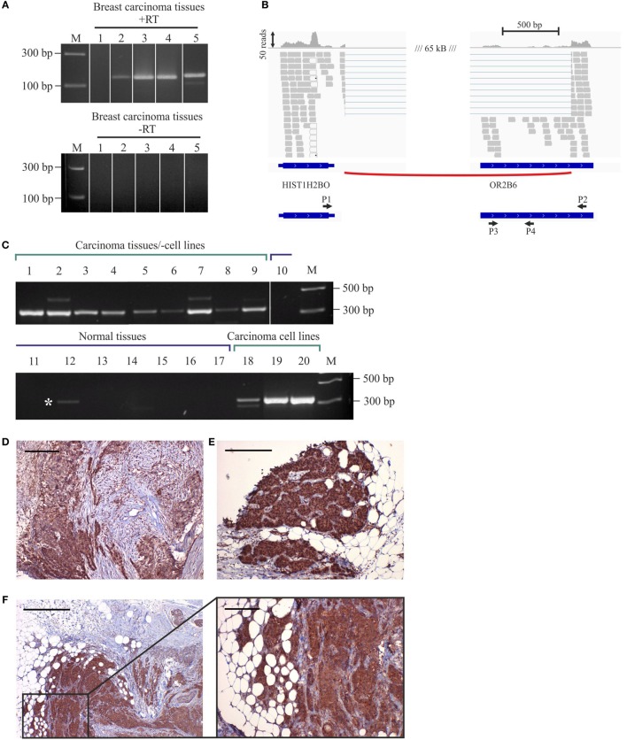
Expression of OR2B6 in human breast carcinoma tissues and detection of fusion transcripts by reverse transcriptase PCR (RT-PCR). (A) Validation of OR2B6 expression in five different breast carcinoma tissues via RT-PCR. +, +RT, cDNA; −, −RT, RNA; M, marker; bp, base pairs. (B) Visualization of the fusion transcript of OR2B6 and HIST1H2BO via the integrative genomic viewer. (C) Validation of the expression of the fusion transcript of OR2B6 and HIST1H2BO via RT-PCR. +, +RT, cDNA; −, −RT, RNA; M, marker; bp, base pairs. (D) Immunhistochemical staining of metaplastic breast carcinoma tissue using specific human α-OR2B6 antibody. Tumor details: G3, triple negative. Shown is the expression of the receptor protein in a triple-negative metaplastic breast carcinoma in G3. Cancer cells are specifically stained, surrounding connective tissue fibers and fat cells show no staining. (E) Immunhistochemical staining of invasive ductal breast carcinoma tissue using specific human α-OR2B6 antibody. Shown is the expression of the receptor in an E+/PR−/HER2− carcinoma in G2 (estrogen 100%: ER+, progesterone 0%: PR−, Her2 negative: HER2−). Protein expression is visualized using DAB chromogenic staining and co-staining with hematoxylin (HE) is used to illustrate the tissue architecture. (F) Immunhistochemical staining of invasive ductal breast carcinoma tissue using specific human α-OR2B6 antibody. Tumor details: G3, triple negative. Shown is the expression of the receptor protein in a triple-negative invasive ductal breast carcinoma in G3. Scale bar: 100 µm.

