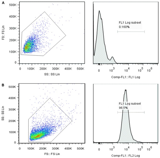
FIGURE 1.

Flow cytometry analysis of the surface maker of the FLSs. Mouse IgG isotypes labeled with FITC served as a negative control (A). The positive rate of 0.16% was chosen as the background (as shown in the box) and then the FITC-labeled VCAM-1 (B) expression rate was examined. The results showed that the expression of VCAM-1 in the cell suspension was 99.5%, indicating that the main component of cultured third-generation synoviocytes was FLSs.
