
Figure 6.
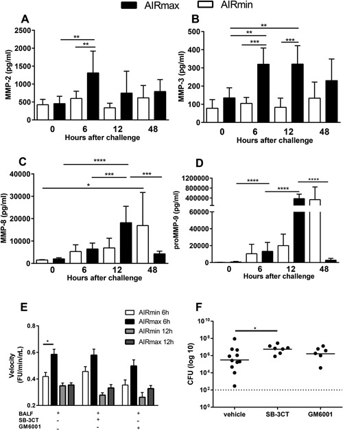
Expression and activity of MMPs, in response to pneumococcal challenge, are impaired in AIRmin mice. Animals (4 to 6 per group) were submitted to intranasal pneumococcal challenge with the ATCC6303 strain. (A–D) Mice were euthanized at different time‐points post‐challenge and BALF were collected for the analysis of MMPs by luminex. Results were expressed by means for each group with SD and are representative of two independent experiments. (E) Enzymatic activity was measured in BALF (from 8 mice per group) collected at 6 and 12 h post‐challenge, using a FRET substrate, Abz‐AGLA‐EDDnp. MMP inhibition was tested by previous incubation of BALF with GM6001 or SB‐3CT. BALF from two independent experiments were tested and reactions were performed in triplicates. Results were expressed by means of each group with SEM. (F) Mice were inoculated with the MMP inhibitors GM6001 or SB‐3CT 2 h before the pneumococcal challenge and 24 h later. Mice were euthanized at 48 h post‐challenge and CFU was evaluated by plating lung macerates on blood‐agar. Graphs were composed with results of two independent experiments. Circles represent each individual and lines represent the medians of the groups. *p < 0.05; **p < 0.01; ***p < 0.001, and ****p< 0.0001 (A–D) Two‐way ANOVA with Tukey's post‐test, (E) Two‐way ANOVA with Tukey's post‐test, (F) Unpaired T test.