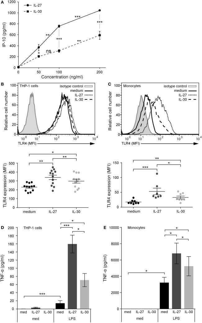
Figure 1.
IL-30 and IL-27 prime cells for enhanced lipopolysaccharide (LPS) responsiveness via TLR4 upregulation. (A) THP-1 cells were treated with IL-27 or IL-30 at 50, 100, or 200 ng/mL for 24 h. IP-10 production was measured in cell-free supernatants by ELISA. Data presented are the mean ± SD of four different THP-1 technical replicate wells. Mann–Whitney U tests were used for statistical analyses between groups as indicated. (B) THP-1 cells and (C) primary human monocytes were stimulated with or without recombinant IL-27 (50 ng/mL) or IL-30 (50 ng/mL) for 24 h. TLR4 expression was measured using flow cytometry, and representative histograms are shown (top). Mean fluorescence intensity corresponding to TLR4 expression was measured for each different THP-1 experiment and different monocyte donors. Data presented include mean ± SEM of all data points (bottom). (D) THP-1 cells and (E) primary human monocytes were treated with or without recombinant IL-27 (50 ng/mL) or IL-30 (50 ng/mL) for 16 h and then washed and stimulated with LPS (1 µg/mL) for 4 h. TNF-α production was measured in cell-free supernatants by ELISA. Data presented are the mean ± SEM of eight different THP-1 experiments or six different monocyte donors. Mann–Whitney U tests were used for statistical analyses between medium and IL-27/IL-30. Wilcoxon matched-pairs signed-rank test was used for statistical analyses between IL-27 and IL-30. *p ≤ 0.05; **p ≤ 0.01; ***p ≤ 0.001.

