Figure 3.
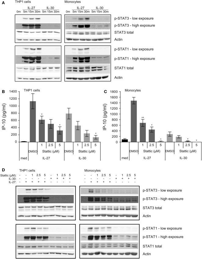
IL-30 and IL-27 differentially activate STAT3 and STAT1. (A) THP-1 cells and primary human monocytes were treated with or without recombinant IL-27 (50 ng/mL) or IL-30 (50 ng/mL) for 5, 15, or 30 min and STAT3 and STAT1 phosphorylation was measured by immunoblot. Blots were stripped and reprobed for total STAT3 and STAT1 levels as well as β-actin as a loading control. Blots are representative of three different THP-1 experiments or three different monocyte donors. (B,D) THP-1 cells and (C,D) primary human monocytes were treated with or without recombinant IL-27 (50 ng/mL) or IL-30 (50 ng/mL) in the presence or absence of Stattic (1–5 µM as indicated) or DMSO solvent control for (B,C) 24 h or (D) 30 min. (B,C) IP-10 production was measured in cell-free supernatants by ELISA. Data presented are the mean ± SEM of three different THP-1 experiments or three different monocyte donors. Mann–Whitney U tests were used for statistical analyses between respective IL-27 or IL-30 control (DMSO) and Stattic treatments. *p ≤ 0.05; **p ≤ 0.01. (D) Phosphorylation of STAT3 and STAT1 were measured by immunoblot. Total STAT3 and STAT1 levels were used as controls, and β-actin was used as a loading control. Blots are representative of three different THP-1 experiments or three different monocyte donors.

