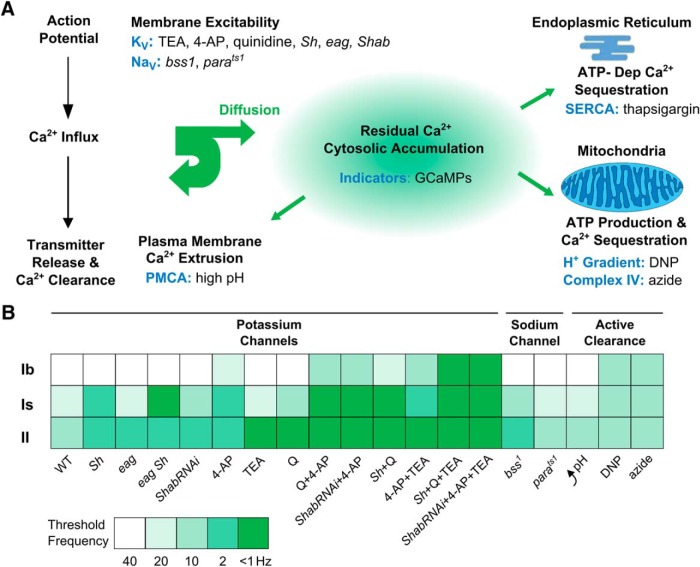
Figure 16.
Presynaptic cytosolic residual Ca2+ regulation in Drosophila NMJ synapses. A, Summary diagram of the relevant cellular mechanisms. The targets of manipulations in this study are shown in blue, including Na+ and K+ channels (NaV and KV), PMCA, and H+ gradient and Complex IV of mitochondria, together with the corresponding mutational and pharmacological manipulations. B, A summary diagram comparing effects of specific experimental manipulations on type Ib, Is, and II synaptic terminals of c164-GCaMP1.3 and nSyb-GCaMP6m. The extent of GCaMP signal enhancement is indicated by reduction in the effective frequencies of stimulation. Threshold frequencies for producing clearly detectable GCaMP signals are color coded. Lower threshold frequencies reflect greater excitability or more hampered Ca2+ clearance. More than 1 Hz indicates the cases where individual stimuli evoked the giant hallmark GCaMP signals due to extreme hyperexcitability (compare Figs. 5, 6). 4-AP, 200 µM; TEA, 10 mM; and Q, 20 µM quinidine.

