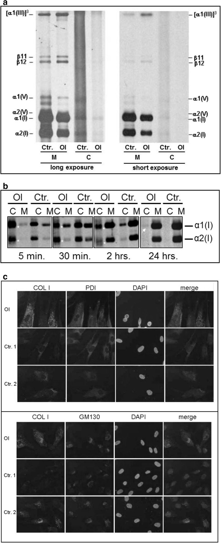
Fig. 2.
Collagen synthesis and secretion in fibroblasts cultures of the OI fetus (OI) and a control (Ctr.); a steady-state analysis showing decreased levels of type I collagen α1(I) and α2(I) relative to type III collagen [α1(III)]3 in the medium (M) of the OI fibroblasts suggesting that less collagen type I was produced and secreted by these cells; b pulse-chase analysis showing a decreased amount of type I collagen chains secreted from the cell layer (C) into the medium layer (M) in the OI fibroblasts compared to the control in the 5-min, 30-min, and 2-h chases. Also, bands with a broader migration pattern are visible in the cell layer (C) of the OI fibroblasts (white asterisk) and a small amount of collagen is still visible after 24-h chase in the cell layer of the OI sample (white arrowhead) but not the control; c Co-immunofluorescent staining of type I collagen (COL I) together with ER (PDI) or Golgi (GM130) markers. DAPI was used to stain nuclei and an overlay of COL I with either PDI or GM130 is given showing increased staining for type I collagen in the OI cells and only partial overlap with ER and Golgi markers

